
Antibacterial and Antifungal Activities of Saponin Extracts from Three Congolese Medicinal Plants of the Fabaceae Family
-
Gedeon M. Siwala
Center for the Study of Natural Substances of Plant Origin, Faculty of Pharmaceutical Sciences, University of Kinshasa, B.P. 212, Kinshasa XI, Democratic Republic of Congo
Carmel G. KandheCenter for the Study of Natural Substances of Plant Origin, Faculty of Pharmaceutical Sciences, University of Kinshasa, B.P. 212, Kinshasa XI, Democratic Republic of Congo
Grace M. MantshumbaCenter for the Study of Natural Substances of Plant Origin, Faculty of Pharmaceutical Sciences, University of Kinshasa, B.P. 212, Kinshasa XI, Democratic Republic of Congo
Brandon N. MikokoCenter for the Study of Natural Substances of Plant Origin, Faculty of Pharmaceutical Sciences, University of Kinshasa, B.P. 212, Kinshasa XI, Democratic Republic of Congo
Cyprien M. LukukulaLaboratory of Experimental and Pharmaceutical Microbiology, Faculty of Pharmaceutical Sciences, University of Kinshasa, Kinshasa, Democratic Republic of Congo
Grégoire V. MbusaLaboratory of Experimental and Pharmaceutical Microbiology, Faculty of Pharmaceutical Sciences, University of Kinshasa, Kinshasa, Democratic Republic of Congo
Michel K. MpuzaCenter for the Study of Natural Substances of Plant Origin, Faculty of Pharmaceutical Sciences, University of Kinshasa, B.P. 212, Kinshasa XI, Democratic Republic of Congo
Isaac E. Kaba
Department of Chemistry, College of Chemistry, University of California, Berkeley, CA 94720, United States
| Received 14 Jun, 2026 |
Accepted 31 Jul, 2026 |
Published 20 Aug, 2026 |
Background and Objective: Antimicrobial resistance is a critical global health threat, particularly in Sub-Saharan Africa, where access to effective treatments is very limited. Saponins from Fabaceae plants exhibit broad pharmacological properties, yet several Congolese species remain pharmacologically unexplored. This study evaluated the antibacterial and antifungal activities of saponin-rich extracts from three Congolese Fabaceae: Millettia laurentii De Wild., Millettia dubia De Wild., and Pentaclethra eetveldeana De Wild. & T. Durand. Materials and Methods: Saponins were extracted from the trunk and root bark of plant species using 80% methanol maceration and reflux, followed by diethyl ether precipitation. A dialyzed fraction of M. dubia was prepared using a 10,000 Da cut-off membrane. Antimicrobial activity was assessed by broth microdilution assay against Staphylococcus aureus ATCC 25923, Escherichia coli ATCC 25922, Pseudomonas aeruginosa ATCC 15442, and Candida kruseiATCC 6258. Results: Extraction yields ranged from 3.27 to 3.6%. All five extracts selectively inhibited P. aeruginosa; the dialyzed M. dubia extract showed the greatest potency (MIC = 15.625 μg/mL). No activity was detected against E. coli or S. aureus. The dialyzed M. dubia extract also demonstrated excellent antifungal activity against C. krusei(MIC = 15.625 μg/mL), outperforming fluconazole. Conclusion: Millettia dubia saponins exhibit potent activity against P. aeruginosa and C. kruseifollowing partial purification, suggesting the antimicrobial potential of this species. Our findings contribute to the valorization of Congolese flora as a potential source of new antimicrobial leads.
| Copyright © 2026 Siwala et al. This is an open-access article distributed under the Creative Commons Attribution License, which permits unrestricted use, distribution, and reproduction in any medium, provided the original work is properly cited. |
INTRODUCTION
Antimicrobial resistance (AMR) is one of the most critical public health threats of the 21st century. Driven by the misuse and overuse of antimicrobials in human medicine, veterinary practice, and agriculture, AMR contributes to millions of deaths annually and is projected to become the leading cause of mortality by 2050 if no decisive action is taken, with associated economic losses estimated in the trillions of dollars1,2. In Africa, the crisis is compounded by inadequate healthcare infrastructure, limited diagnostics, and restricted access to a new generation of antibiotics3.
In many rural communities of the Democratic Republic of Congo (DRC) and other African nations, more than 80% of the population relies primarily on traditional herbal remedies for primary healthcare4,5. This dependence on traditional plant-based medicines, although born of necessity, has long fueled scientific interest in the pharmacological potential of African medicinal plants. In recent decades, renewed efforts have been directed toward systematically characterizing the phytochemical profiles and biological activities of plant secondary metabolites, including phenolic compounds, alkaloids, terpenes, essential oils, and saponins6-9. Among these metabolites, saponins have attracted particular attention because of their structural diversity, amphiphilic nature, and broad range of pharmacological properties, which include anti-inflammatory, antidiabetic, anticancer, antiviral, and antimicrobial activities10,11. Saponins are naturally occurring glycosides composed of a lipophilic aglycone linked to one or more hydrophilic sugar chains. Their amphiphilic character confers strong surfactant properties, manifested as stable foam formation in aqueous solutions, a key feature used in preliminary phytochemical screening12,13. Their ability to interact with membrane sterols and disrupt lipid bilayer integrity underlies both their hemolytic properties and their antimicrobial potential14.
The Fabaceae family is among the plant families richest in saponins, encompassing numerous genera with demonstrated pharmacological relevance, including Millettia and Pentaclethra15,16. Millettia laurentii De Wild., commonly known as Wengé, is a large tropical tree native to the Congo Basin and is listed as an Endangered species by the IUCN due to habitat destruction and overexploitation. Its bark has traditionally been used in the DRC in aqueous extractions for the treatment of convulsive cough and asthma, and has been reported to possess antitumoral, anti-inflammatory, and bactericidal activities17,18. Millettia dubia De Wild. is a liana or small tree endemic to the DRC, locally known as Bonkelele in Lingala (a national language in DRC), for which no prior pharmacological data have been reported. On the other hand, Pentaclethra eetveldeana De Wild. & T. Durand, which is a tree of the African tropical forest distributed from Gabon to Cabinda, is traditionally used in the Republic of Congo and DRC for respiratory diseases, constipation, gastritis, helminthiasis, and hemorrhoids19-22. Triterpenoid saponins have been previously isolated and characterized from the trunk bark20. Despite the ethnomedical importance of these species, no comparative antimicrobial study has been conducted on all three simultaneously, and M. dubia remains entirely unexplored pharmacologically. This study, therefore, aimed to evaluate the saponin content of different plant organs using the foam test; extract and quantify saponin-rich fractions from the most saponin-rich parts; assess the effect of dialysis purification on the antimicrobial activity of M. dubia extracts; and evaluate the antibacterial and antifungal activities of all extracts against four clinically relevant microbial strains.
MATERIALS AND METHODS
Study area and duration: Plant material was collected from the campus of the University of Kinshasa (UNIKIN), located in the Commune of Lemba, Kinshasa, Democratic Republic of Congo (4°26 S, 15°19 E). The University of Kinshasa campus is situated in the Kinshasa Plateau Region, characterized by a tropical savanna climate (Köppen Aw) with a mean annual temperature of approximately 25°C and a mean annual rainfall of 1,400 mm, distributed across two rainy seasons (September-December and February-May) and two dry seasons. The campus hosts a diverse collection of indigenous and naturalized woody species, making it a well-established site for the botanical collection of Congolese flora. The study was conducted from January 2023 to March 2024. Bark specimens (M. laurentii trunk bark, M. dubia root and trunk bark, and P. eetveldeana trunk and root bark) were collected in January 2023. Bark collection is feasible year-round and is not constrained by a specific phenological window. Seeds of M. laurentii were harvested in May 2023, corresponding to the flowering and fruiting period of the species.
|
| Table 1: | Culture conditions used for each microbial strain | |||
| Microbial strain | Culture medium | Incubation temperature (°C) |
| Escherichia coli ATCC 25922 | MacConkey agar | 35±2 |
| Pseudomonas aeruginosa ATCC 15442 | Cetrimide agar | 30 |
| Staphylococcus aureus ATCC 25923 | Mannitol salt agar | 35±2 (aerobic) |
| Candida krusei ATCC 6258 | Sabouraud dextrose agar | 35-37 |
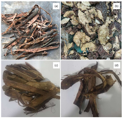
Plant material: Millettia laurentii De Wild., Millettia dubia De Wild., and Pentaclethra eetveldeana De Wild. & T. Durand were collected from the University of Kinshasa campus. Bark specimens (trunk and root) were air-dried at ambient temperature (Fig. 1) and ground into a fine powder. All species were authenticated by Mr. Boniface Lukebakio Nlandu of Institut National d’Etudes et de Recherches Agronomiques (INERA Herbarium, University of Kinshasa). Voucher specimens were deposited under reference numbers GS001 (M. laurentii trunk bark), GS002 (P. eetveldeana trunk bark), GS003 (P. eetveldeana root bark), GS004 (M. dubia trunk bark), and GS005 (M. dubia root bark).
Chemicals, reagents, and microbial strains: Distilled water, 80% methanol, and diethyl ether were used for all extractions. Dimethyl sulfoxide (DMSO, 5%), tryptic soy broth (TSB), Mueller-Hinton broth (MHB), resazurin sodium salt, phosphate-buffered saline (PBS), and standard microbiological media were of analytical grade. Ciprofloxacin and fluconazole (Sigma-Aldrich) served as positive controls. Reference microbial strains, including Staphylococcus aureus ATCC® 25923™, Escherichia coli ATCC® 25922™, Pseudomonas aeruginosa ATCC® 15442™, and Candida krusei ATCC® 6258™, were supplied by the Microbiology Laboratory, Faculty of Pharmaceutical Sciences, University of Kinshasa. Culture conditions are described in Table 1.
Foam test: Two grams of dried powder were macerated in 30 mL of distilled water at ambient temperature for 24 hrs, heated at 70°C for 90 min, and filtered. The filtrate was vigorously shaken for 5 min in a 15-cm test tube, and the foam column height was measured immediately and at 15 and 30 min. Results were scored as: (++) strongly positive (column >10 cm, stable after 15 min); (+) positive (column >10 cm, gradual degradation); (0) weakly positive (<10 cm); and (–) negative.23
Saponin extraction: Dried plant powder was macerated in 500 mL of 80% methanol for 6 days, then subjected to reflux extraction at 80°C for 3 hrs24. After filtration, the extract was concentrated by evaporation and further reduced under vacuum to approximately 120 mL. Saponins were precipitated by the addition of three volumes of diethyl ether, collected by Büchner filtration, and dried under vacuum.
Dialysis purification of M. dubia extract: One gram of crude M. dubia saponin extract was dissolved in 100 mL of distilled water and transferred to a pre-soaked cellophane membrane (10,000 Da cut-off, Amicon). Dialysis was performed against 500 mL of distilled water with gentle agitation for 24 hrs, with water replaced every 4 hrs25. The retentate was recovered and freeze-dried. A loss of 320 mg (32%) was recorded, yielding 680 mg of dialyzed extract.
Antimicrobial activity (microdilution assay): Antimicrobial activity was evaluated by broth microdilution in 96-well polystyrene microplates according to CLSI guidelines26. Each extract was dissolved in 5% DMSO and diluted to 4000 μg/mL in TSB. Two-fold serial dilutions yielded final concentrations of 2000-15.625 μg/mL per well (200 μL total volume per well). Growth and sterility controls were included on each plate. After incubation at appropriate temperatures (35±2°C for S. aureus and E. coli; 35±2°C for P. aeruginosa; 35°C for C. krusei), 10 μL of resazurin (0.01%, w/v) was added, and plates were re-incubated for 4 hrs. The MIC was the lowest concentration, showing no color change from blue to pink27. Experiments were performed in triplicate across three independent sessions.
RESULTS AND DISCUSSION
Foam test and saponin distribution: The foam test results are presented in Table 2. A strongly positive result (++) was obtained from the trunk bark of M. laurentii (column height 11 cm, stable after 30 min) and root bark of M. dubia (11 cm). In contrast, seeds of M. laurentii and trunk bark of M. dubia showed negative or weakly positive results, respectively. Both trunk and root bark of P. eetveldeana showed positive responses (+), with column heights of 5 and 4 cm, respectively. These results indicate preferential accumulation of saponins in bark organs, consistent with reports from other Fabaceae14. The findings guided the selection of plant material: Trunk bark for M. laurentii, root bark for M. dubia, and both bark types for P. eetveldeana, in agreement with prior phytochemical characterizations20.
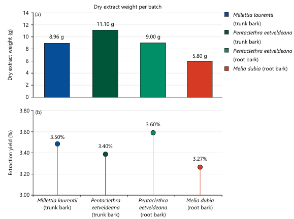
Saponin extraction yields: Extraction yields ranged from 3.27 to 3.6%, corresponding to dry extract masses of 5.8-11.1 g per batch (Fig. 2a-b). The highest absolute yield was obtained from P. eetveldeana trunk bark (11.1 g; 3.4%), while P. eetveldeana root bark gave the highest percentage yield (3.6%). Millettia. laurentii trunk bark yielded 8.96 g (3.5%), and M. dubia root bark yielded 5.8 g (3.27%). All extracts appeared as amorphous brownish or golden residues, consistent with saponin-enriched fractions24. These yields fall within the range commonly reported for crude saponin fractions obtained by methanol maceration and precipitation10. It is important to note that saponin content was not quantified spectrophotometrically in the crude extracts; yields are reported as gravimetric extraction yields (%), which reflect total precipitable material rather than pure saponin mass. Additionally, the hemolytic activity was not assessed for any extract, including the dialyzed M. dubia fraction. Given that saponins are classically hemolytic due to their interaction with membrane cholesterol, the absence of hemolysis data may limit the interpretation of the selectivity and safety of the active fractions. Future work should incorporate quantitative saponin determination, for example, using a vanillin-sulfuric acid or β-sitosterol calibration method, to allow accurate inter-species and inter-organ comparisons of saponin loading.
|
| Table 2: | Foam column height and stability of plant organs from three Congolese Fabaceae species as indicators of saponin content | |||
| Plant species | Organ | Column height (cm) | Structure after 15 min | Structure after 30 min | Score |
| Millettia laurentii | Trunk bark | 11 | Intact | Very slightly modified | ++ |
| Millettia laurentii | Seeds | 1.5 | Intact | Intact | – |
| Millettia dubia | Trunk bark | 5 | 1.0 cm | Gradually collapses | 0 |
| Millettia dubia | Root bark | 11 | 1.9 cm | Tends to remain stable | ++ |
| Pentaclethra eetveldeana | Trunk bark | 5 | 1.0 cm | Tends to remain stable | + |
| Pentaclethra eetveldeana | Root bark | 4 | 1.9 cm | Tends to remain stable | + |
| (++): Strongly positive, (+): Positive, (0): Weakly positive and (–): Negative | |||||
Antibacterial activity: The MIC values for all extracts are presented in Table 3. All five extracts demonstrated selective inhibition of P. aeruginosa, while no activity was detected against E. coli or S. aureus at concentrations up to 2000 μg/mL. Against P. aeruginosa, the dialyzed M. dubia extract was by far the most potent (MIC = 15.625 μg/mL), eight-fold lower than its crude counterpart (MIC = 125 μg/mL). Pentaclethra eetveldeana root bark showed the next best activity (MIC = 31.250 μg/mL), followed by M. laurentii trunk bark (MIC = 62.5 μg/mL). Although these values remain higher than those of ciprofloxacin (MIC = 2.5 μg/mL), they are consistent with values reported for crude saponin fractions from other species28. The resistance of E. coli and S. aureus is consistent with observations that the antibacterial activity of bidesmosidic saponins becomes apparent only after hydrolysis to more active monodesmosidic forms29. Both the aglycone nature and sugar chain composition critically influence membrane disruption capacity12,30,31. The selective activity against P. aeruginosa may relate to specific membrane composition differences, which make this bacterium more vulnerable to saponin-mediated disruption14.
| Table 3: | Minimum inhibitory concentrations (MICs, μg/mL) of saponin-rich extracts and controls against tested microbial strains | |||
| Pathogen | Millettia dubi (EHAER) | Millettia dubi Dialyzed | Millettia laurentii (EHAET) | Pentaclethra eetveldeana (EHAER) | Pentaclethra eetveldeana (EHAET) | Control |
| Pseudomonas aeruginosa | 125 | 15.625 | 62.5 | 31.25 | 125 | 2.5 (CIP) |
| Escherichia coli | >2000 | >2000 | >2000 | >2000 | >2000 | 2.0 (CIP) |
| Staphylococcus aureus | >2000 | >2000 | >2000 | >2000 | >2000 | 5.0 (CIP) |
| Candida krusei | 2000 | 15.625 | 1000 | 250 | 1000 | 2000 (FLU) |
| EHAER: Hydroalcoholic extract of root bark, EHAET: Hydroalcoholic extract of trunk bark, CIP: Ciprofloxacin, FLU: Fluconazole. Values >2000 indicate no inhibitory activity at the highest tested concentration. All experiments were performed in triplicate across three independent sessions | ||||||
Antifungal activity: The dialyzed M. dubia extract demonstrated exceptional antifungal potency (MIC = 15.625 μg/mL), lower than the fluconazole control (MIC = 2000 μg/mL). Pentaclethra eetveldeana root bark also showed appreciable antifungal activity (MIC = 250 μg/mL), while M. laurentii and crude M. dubia showed weaker effects (MIC = 1000 and 2000 μg/mL, respectively). The superior antifungal activity reflects saponin enrichment and removal of interfering low-molecular-weight compounds during dialysis. Saponins are known to disrupt fungal membranes by interacting with ergosterol, forming pores that compromise membrane integrity14,32. This mechanism explains the stronger antifungal relative to antibacterial activity, as fungal membranes are sterol-rich while bacterial (particularly Gram-negative) membranes typically lack or contain different sterols. These findings are consistent with potent antifungal activities reported for triterpenoid saponins from other organisms33-35.
Effect of dialysis purification on antimicrobial activity: Dialysis purification dramatically enhanced M. dubia extract activity: MIC against P. aeruginosa dropped from 125 to 15.625 μg/mL (8-fold); MIC against C. krusei dropped from 2000 to 15.625 μg/mL (128-fold). This improvement reflects two complementary effects: Removal of low-molecular-weight impurities (sugars, salts, organic acids) that compete with saponins in membrane interactions, and enrichment in saponin concentration per unit mass. Comparable purification-dependent improvements have been reported by Hassan et al.30 for saponin fractions from guar, quillaja, yucca, and soybean. Stuardo and Martín25 similarly showed that partial alkaline hydrolysis of quinoa saponins dramatically improved antifungal activity, mechanistically analogous to the enrichment achieved here. Our results indicate that the antimicrobial potential of M. dubia saponins may be substantially underestimated when crude extracts alone are tested, and that individual saponin compounds are likely to have even lower MIC values than 15.625 μg/mL. Further work, including isolation of individual saponin compounds by chromatographic fractionation, is necessary to confirm the observed activity and better determine structure-activity relationships.
CONCLUSION
This study constitutes a comparative evaluation of the antimicrobial activities of saponin-rich extracts from Millettia laurentii, Millettia dubia, and Pentaclethra eetveldeana from the Democratic Republic of Congo. All five extracts selectively inhibited Pseudomonas aeruginosa, while no activity was detected against E. coli or S. aureus. Most importantly, dialyzed M. dubia saponins demonstrated outstanding activity against both P. aeruginosa and C. krusei, outperforming fluconazole, consistent with the antimicrobial potential of individual saponin constituents recently isolated from this species by preparative HPLC, NMR spectroscopy, and mass spectrometry. These findings identify M. dubia as a priority candidate for phytochemical drug discovery targeting resistant P. aeruginosa and C. krusei infections and support the valorization of Congolese Fabaceae as a source of novel antimicrobial leads. Future work should focus on the evaluation of dialyzed fractions from M. laurentii and P. eetveldeana to determine whether purification equally enhances their activities; mechanistic studies to elucidate the mode of action of the active saponins against P. aeruginosa and C. krusei; and cytotoxicity and in vivo pharmacological evaluation to establish the safety profiles required for preclinical development.
AUTHOR CONTRIBUTIONS
G.M. Siwala: Conceptualization, Methodology, Investigation, Writing (original draft). C.G. Kandhe: Formal Analysis, Data Curation, Writing (review and editing). G.M. Mantshumba: Investigation, Data Curation. B.N. Mikoko: Investigation, Data Curation. C.M. Lukukula: Investigation, Validation. G.V. Mbusa: Investigation, Validation. M.K. Mpuza: Conceptualization, Supervision, Project Administration. I.E. Kaba: Supervision, Investigation, Resources, Data Curation, Visualization, Conceptualization, Writing (original draft; review and editing).
CONFLICT OF INTEREST
The authors declare no conflict of interest.
FUNDING
The research did not receive any specific grant from funding agencies.
SIGNIFICANCE STATEMENT
This study highlights the potential of saponin-rich bark extracts from selected Congolese Fabaceae species as promising sources of antimicrobial agents. The observed selective activity against Pseudomonas aeruginosa, together with the enhanced potency following dialysis, particularly in Millettia dubia, demonstrates the importance of extract purification in revealing strong bioactive compounds. The superior antifungal performance of dialyzed M. dubia compared to fluconazole further supports its therapeutic relevance. These findings identify M. dubia as a high-priority candidate for future phytochemical isolation and development of novel agents targeting resistant bacterial and fungal pathogens.
ACKNOWLEDGMENTS
The authors gratefully acknowledge Mr. Boniface Lukebakio Nlandu of the National Institute of Agronomic Studies and Research (INERA Herbarium, University of Kinshasa) for botanical authentication. The authors thank the staff of the Center for the Study of Natural Substances of Plant Origin (CESNOV) and the Laboratory of Experimental and Pharmaceutical Microbiology of the University of Kinshasa for providing microbial reference strains and technical assistance.
REFERENCES
- Murray, C.J.L., K.S. Ikuta, F. Sharara, L. Swetschinski and G.R. Aguilar et al., 2022. Global burden of bacterial antimicrobial resistance in 2019: A systematic analysis. Lancet, 399: 629-655 .
- O'Neill, J., 2016. Tackling Drug-Resistant Infections Globally: Final Report and Recommendations. Review on Antimicrobial Resistance, London, United Kingdom, Pages: 80.
- WHO, 2023. Antimicrobial Resistance. World Health Organization, Geneva, Switzerland.
- WHO, 2019. WHO Global Report on Traditional and Complementary Medicine 2019. World Health Organization, Geneva, Switzerland, ISBN: 9789241515436, Pages: 226.
- Mutombo, C.S., S.A. Bakari, V.N. Ntabaza, A. Nachtergael, J.B.S. Lumbu, P. Duez and J.B. Kahumba, 2022. Perceptions and use of traditional African medicine in Lubumbashi, Haut-Katanga Province (DR Congo): A cross-sectional study. PLoS ONE, 17.
- Stapleton, P.D., S. Shah, J.M. Hamilton-Miller, Y. Hara, Y. Nagaoka, A. Kumagai and P.W. Taylor, 2004. Anti-Staphylococcus aureus activity and oxacillin resistance modulating capacity of 3-O-acyl-catechins. Int. J. Antimicrob. Agents., 24: 374-380.
- Elaka, I.K., P.M. Kapepula, N.K. Ngombe, J.M. Mpoyi and D.M. Lukusa et al., 2020. Microscopic features, antioxidant and antibacterial capacities of plants of the congolese cosmetopoeia, raw materials of cosmeceuticals. J. Biosci. Med., 8: 149-166.
- Muipata, M.M., S.N. Mbwaka, P.M. Mbombo, I.E. Kaba and J.M. Mukanya et al., 2025. Phytochemical analysis, antioxidant and antibacterial capacities of a congolese traditional recipe and medicinal plants commonly used in prostate diseases. J. Biosci. Med., 13: 382-399.
- Kaba, I.E., C.G. Kandhe, M.M. Muipata, B.P.I. Muntokole and J.M. Mukanya et al., 2026. Ethnopharmacological survey, fumigation efficiency, and pharmacovigilance of plant-based medicines for respiratory disease during COVID-19 in the Democratic Republic of Congo. Res. Square.
- Augustin, J.M., V. Kuzina, S.B. Andersen and S. Bak, 2011. Molecular activities, biosynthesis and evolution of triterpenoid saponins. Phytochemistry, 72: 435-457.
- Piao, X., H. Zhang, J.P. Kang, D.U. Yang and Y. Li et al., 2020. Advances in saponin diversity of Panax ginseng. Molecules, 25.
- Vincken, J.P., L. Heng, A. de Groot and H. Gruppen, 2007. Saponins, classification and occurrence in the plant kingdom. Phytochemistry, 68: 275-297.
- Faizal, A. and D. Geelen, 2013. Saponins and their role in biological processes in plants. Phytochem. Rev., 12: 877-893.
- Wink, M., 2015. Modes of action of herbal medicines and plant secondary metabolites. Medicines, 2: 251-286.
- Banzouzi, J.T., A. Prost, M. Rajemiarimiraho and P. Ongoka, 2008. Traditional uses of the African Millettia species (Fabaceae). Int. J. Bot., 4: 406-420.
- Bouzier, A., J. Rojas, S.K.K. Ibinga, A. Lamarti, P. Martin and M. Morillo, 2023. The impact of saponins on health-review. Biointerface Res. Appl. Chem., 13.
- Mofor, C.T., C.R.S. Dontsa, R. Fokom, V.P. Beng and P.H.A. Zollo, 2013. Antifungal and antioxidant activity of crude extracts of three medicinal plants from Cameroon pharmacopea. J. Med. Plants Res., 7: 1537-1542.
- Pertuit, D., A.C. Mitaine-Offer, T. Miyamoto, C. Tanaka and C. Delaude et al., 2022. Oleanane-type glycosides isolated from the trunk barks of the Central African tree Millettia laurentii. Fitoterapia, 159.
- Bouquet, A., 1969. Fetish Priests and Traditional Medicines of Congo (Brazzaville) [In French]. Office for Overseas Scientific and Technical Research, Paris, France, Pages: 282.
- Pertuit, D., M. Kapundu, A.C. Mitaine-Offer, T. Miyamoto, C. Tanaka, C. Delaude and M.A. Lacaille-Dubois, 2019. Triterpenoid saponins from the stem bark of Pentaclethra eetveldeana. Nat. Prod. Commun., 14.
- N’goka, V., S.L.O. Liabagui, C.S. Obiang, H. Begouabe and G.F.N. Ntandou et al., 2023. Pentaclethra eetveldeana leaves from four Congo-Brazzaville Regions: Antioxidant capacity, anti-inflammatory activity and proportional accumulation of phytochemicals. Plants, 12.
- Matala. G.I. and C.A. Lubini, 2019. Environmental and economic potential of a natural regeneration of Pentaclethra eetveldeana in Kikola in Kongo Central / DRC. Int. J. Latest Res. Humanit. Social Sci., 2: 61-73.
- Thakur, M., M.F. Melzig, H. Fuchs and A. Weng, 2011. Chemistry and pharmacology of saponins: Special focus on cytotoxic properties. Botanics: Targets Ther., 1: 19-29.
- Majinda, R.R.T., 2012. Extraction and Isolation of Saponins. In: Natural Products Isolation, Sarker, S.D. and L. Nahar (Eds.), Humana Press, Totowa, New Jersey, ISBN: 978-1-61779-624-1, pp: 415-426.
- Stuardo, M. and R.S. Martin, 2008. Antifungal properties of quinoa (Chenopodium quinoa Willd) alkali treated saponins against Botrytis cinerea. Ind. Crops Prod., 27: 296-302.
- Weinstein, M.P., 2018. Methods for Dilution Antimicrobial Susceptibility Tests for Bacteria that Grow Aerobically. 11th Edn., Clinical Laboratory Standards Institute, United States, ISBN: 9781562388362, Pages: 92.
- Patil, S., S.T. Mohite, S.A. Kulkarni and U.S. Udgaonkar, 2014. Resazurin tube method: Rapid, simple, and inexpensive method for detection of drug resistance in the clinical isolates of Mycobacterium tuberculosis. J. Global Infect. Dis., 6: 151-156.
- Mandal, P., S.P.S. Babu and N.C. Mandal, 2005. Antimicrobial activity of saponins from Acacia auriculiformis. Fitoterapia, 76: 462-465.
- Avato, P., R. Bucci, A. Tava, C. Vitali, A. Rosato, Z. Bialy and M. Jurzysta, 2006. Antimicrobial activity of saponins from Medicago sp.: Structure-activity relationship. Phytother. Res., 20: 454-457.
- Hassan, S.M., J.A. Byrd, A.L. Cartwright and C.A. Bailey, 2010. Hemolytic and antimicrobial activities differ among saponin-rich extracts from guar, quillaja, yucca and soybean. Appl. Biochem. Biotechnol., 162: 1008-1017.
- Li, J. and V. Monje-Galvan, 2023. In vitro and in silico studies of antimicrobial saponins: A review. Processes, 11.
- Guillaume, D. and Z. Charrouf, 2005. Saponins and secondary metabolites of the argan tree (Argania spinosa) [In French]. Cah. Agric., 14: 509-516.
- Bahrami, Y., W. Zhang and C.M.M. Franco, 2018. Distribution of saponins in the sea cucumber Holothuria lessoni; the body wall versus the viscera, and their biological activities. Mar. Drugs, 16.
- Tagousop, C.N., J.D.D. Tamokou, L.D.T. Feugap, D. Harakat, L. Voutquenne-Nazabadioko and D. Ngnokam, 2021. New hemisynthetic oleanane saponin with antimicrobial activities. Adv. Biol. Chem., 11.
- Arabski, M., A. Węgierek-Ciuk, G. Czerwonka, A. Lankoff and W. Kaca, 2012. Effects of saponins against clinical E. coli strains and eukaryotic cell line. J. Biomed. Biotechnol., 2012.
How to Cite this paper?
APA-7 Style
Siwala, G. M., Kandhe, C. G., Mantshumba, G. M., Mikoko, B. N., Lukukula, C. M., Mbusa, G. V., Mpuza, M. K., & Kaba, I. E. (2026). Antibacterial and antifungal activities of saponin extracts from three congolese medicinal plants of the fabaceae family. Research Journal of Medicinal Plants, 20(1), 28–36. https://doi.org/10.3923/rjmp.2026.28.36
ACS Style
Siwala, G. M.; Kandhe, C. G.; Mantshumba, G. M.; Mikoko, B. N.; Lukukula, C. M.; Mbusa, G. V.; Mpuza, M. K.; Kaba, I. E. Antibacterial and Antifungal Activities of Saponin Extracts from Three Congolese Medicinal Plants of the Fabaceae Family. Res. J. Med. Plants 2026, 20 (1), 28–36. https://doi.org/10.3923/rjmp.2026.28.36
AMA Style
Siwala GM, Kandhe CG, Mantshumba GM, et al. Antibacterial and antifungal activities of saponin extracts from three congolese medicinal plants of the fabaceae family. Research Journal of Medicinal Plants. 2026;20(1):28–36. doi:10.3923/rjmp.2026.28.36
Chicago/Turabian Style
Siwala, Gedeon M., Carmel G. Kandhe, Grace M. Mantshumba, et al. 2026. “Antibacterial and Antifungal Activities of Saponin Extracts from Three Congolese Medicinal Plants of the Fabaceae Family.” Research Journal of Medicinal Plants 20 (1): 28–36. https://doi.org/10.3923/rjmp.2026.28.36

This work is licensed under a Creative Commons Attribution 4.0 International License.

